CrystalVue™ is an advanced volume rendering technology that enhances visualization of both internal and external structures in a single rendered image using a unique combination of intensity, gradient and position.
CrystalVue Flow™ is a volume rendering technology that provides additional information of blood flow morphology based on the CrystalVue™ features that visualizes the internal structures by projecting the 3D data, providing better understanding in the anatomic structures and surrounding vascular vessels.
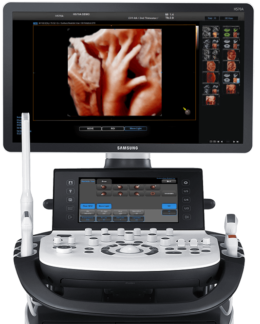
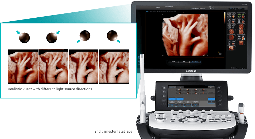
Realistic Vue™ displays high resolution 3D anatomy with details and realistic depth perception. User selectable light source direction creates intricately graduated shadows for better defined anatomical structures.
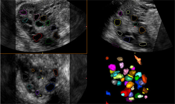
Sonographic parameters have been proven to be effective indicators when assessing in-vitro fertilization (IVF). 5D Follicle™ measures the size and the status of each follicle to provide useful diagnosis information.
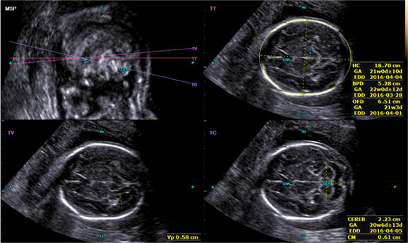
5D CNS™ offers 6 measurements (BPD, HC, OFD, Vp, TCD, CM) from 3 transverse planes of a fetal brain which are the key indicators for intuitive fetal brain visualization. It improves throughput with only a 2 click peration.
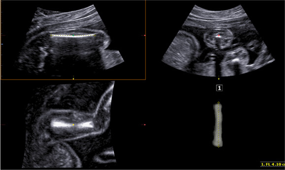
5D LB™ allows easy detection and measurement of fetal long bones from volume data, with intuitive visualization of the fetal structures, resulting in more accurate, yet shorter exam-timed evaluation of fetal condition.
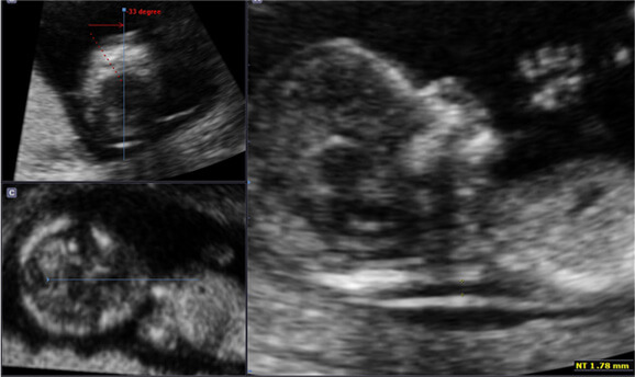
Operator dependency is reduced for the first trimester fetal nuchal translucency measurements with NT measurement solutions. 5D NT™ applies Realistic Vue™ to the automatically detected mid-sagittal view for intuitive confirmation.
A semi-automatic technology for biometric measurement, BiometryAssist™, enables users to measure the fetal growth parameters such as BPD, HC, AC and FL with one click while maintaining exam consistency.
IOTA-ADNEX (International Ovarian Tumor Analysis-Assessment of Different NEoplasias in the adnexa) applies ADNEX* model, an ovarian tumor classification solution of IOTA** Group, to the system, and can perform all procedures from the initial scan to the final report in the ultrasound diagnosis system. When two ultrasound prediction factors are measured, the measurements are automatically populated and the classification result from the 5-step ADNEX model for ovarian tumors is provided.
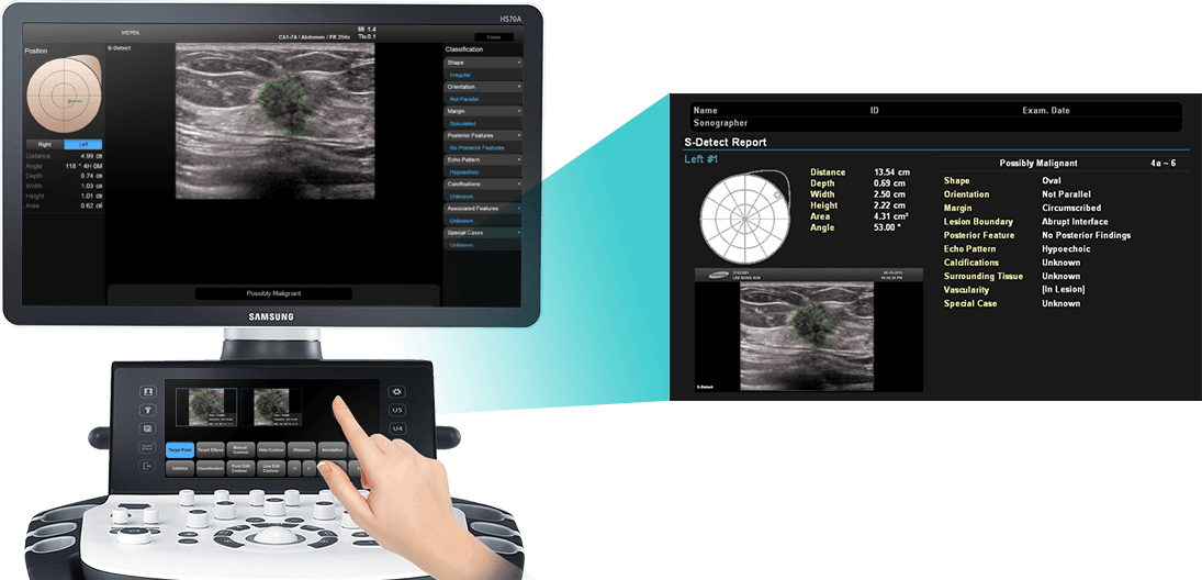
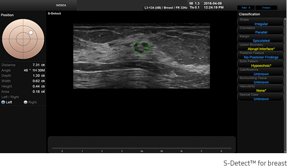
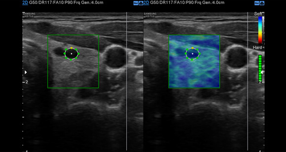
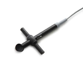
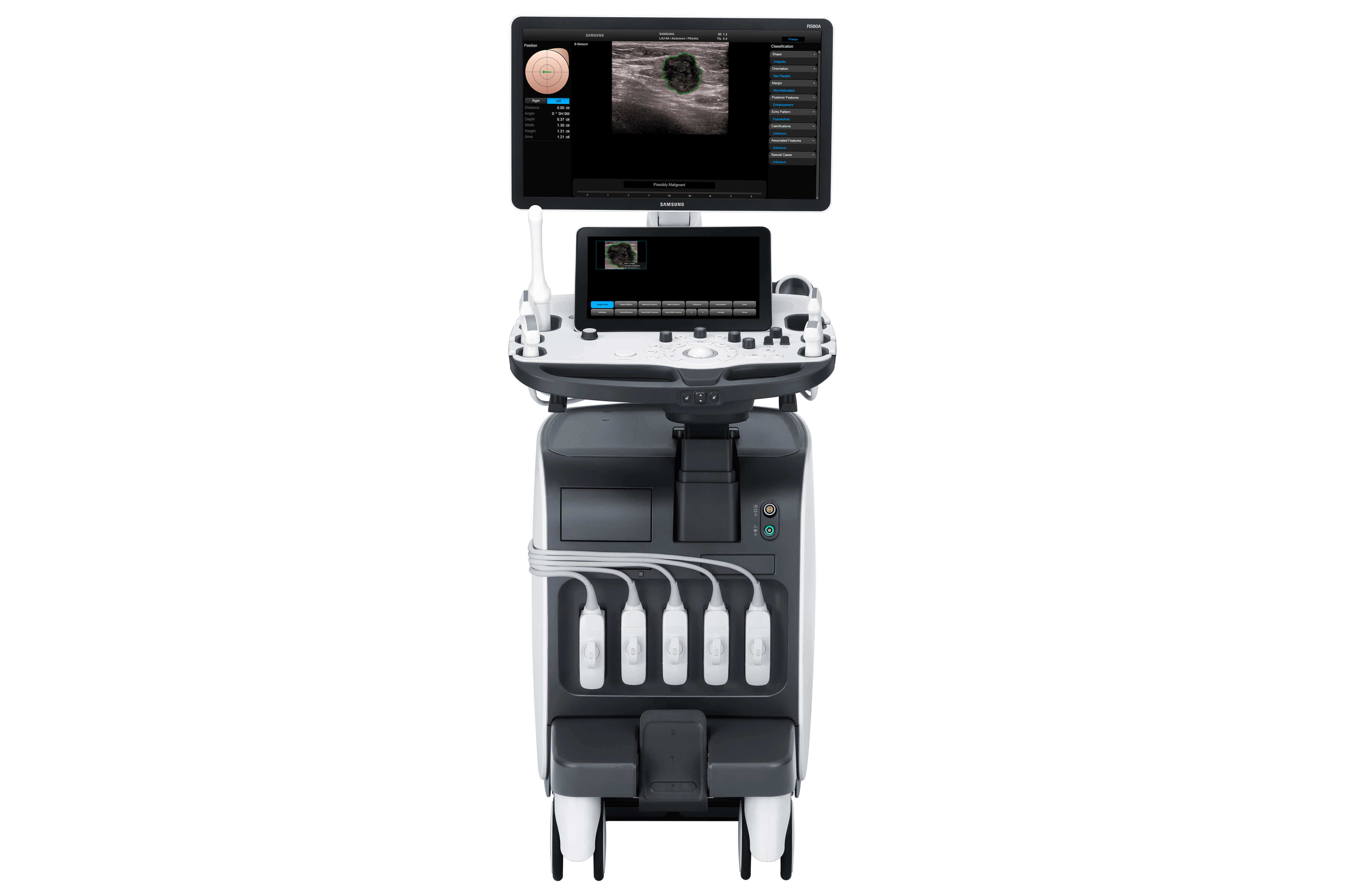
By simply clicking the suspected area, S-Detect™ for Breast draws the lesion area and provides the characteristics of the lesion and a recommendation on whether the lesion is benign or malignant.
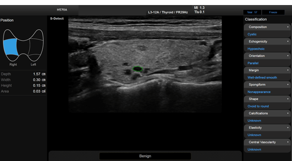
S-Detect™ for Thyroid uses advanced methods based on *K-TIRADS, RUSS and ATA draft to detect and classify suspicious thyroid lesions semi-automatically. This state-of-the art technology helps diagnose patients with confidence and ease, providing accurate, consistent results with an automatic reporting feature.
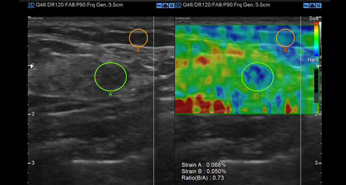
E-Strain™ enables quick and easy calculation of the strain ratio between two regions of interest in day-to-day procedures such as breast, prostate or gynecological examinations.
E-Thyroid™ uses pulsations from the adjacent Carotid Artery and provides an assessment of thyroid lesions.
With the S-VisionTM imaging engine built into RS85, the digital signals produce clear, detailed resolution and tissue uniformity for various types of applications in general imaging.
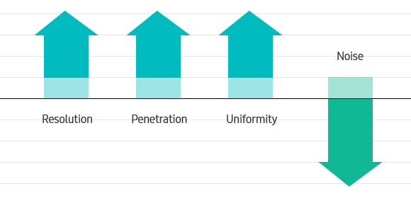
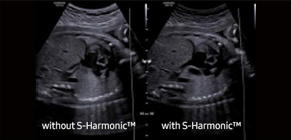
This new harmonic technology makes a clearer image- near to far. Reducing signal noise,S-Harmonic™ provides more uniform ultrasound images.
ClearVision offers speckle reduction, edge enhancement and contrast enhancement for clear and natural images. In addition, ClearVision provides application-specific optimization in live scan mode.

Reach us via email or phone!